Mechanical Scanning Methods
Recording of ultrasound B-images of a simple specimen with two ultrasound probes of different frequencies using a computer-controlled scanner
Using a computer-controlled scanner, ultrasound B-images of a simple specimen are recorded using two ultrasound probes of different frequencies. The image quality of the B-images is analyzed with regard to focus zone, resolution and possible artifacts.
Keywords: Ultrasound echography, A-image, B-image, resolution, image artifacts
To obtain a B-image with an ultrasound transducer, the transducer or the sound beam must be moved along the desired cutting line. Compared to a hand-held scanning procedure, mechanical and electronic scanning methods offer better image quality due to good resolution and freely selectable line density. Due to the low frame rate, however, electronic multi-element scanners are used for real-time images and moving structures. By using ultrasound probes of different frequencies in combination with mechanically guided uniform scanning, the lateral frequency-dependent resolution can be examined and evaluated in the experiment in addition to the axial resolution.
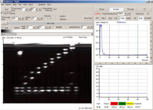
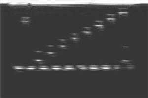
The figure shows the B-scan of an acrylic block with holes of different sizes and arrangements, recorded with a 2 MHz probe. By examining it in a water bath, with the holes filled with water, echoes from both the upper and lower edges of the holes can be seen. The acoustic shadows of the holes above are visible in the bottom echo.
SCOPE OF DELIVERY:
| Item No. | Designation |
|---|---|
| 10400 | Ultrasonic echoscope GS200 |
| 10151 | Ultrasonic probes 1 MHz |
| 10152 | Ultrasonic probes 2 MHz |
| 10201 | Test block (transparent) |
| 10204 | – optional: test block (black) |
| 60200 | CT scanner |
| 60210 | CT control |
| 60120 | CT measuring tank |
| 70200 | Ultrasound gel |
ADDITIONAL EXPERIMENTS:
| PHY08 | Ultrasound B-scan |
| PHY10 | Sound field characteristics |
| PHY20 | Determination of the focus zone |
| IND08 | Defect testing |
| MED02 | Ultrasound examinations on the breast model (mammography) |


