


Basics of Doppler Sonography
Development of the physical and signal theoretical principles for blood flow investigations using the ultrasound Doppler method (Doppler sonography)
The experiment will develop the physical and signal-theoretical principles necessary for blood flow studies using the ultrasound Doppler method. The dependence of the color-coded Doppler spectra on signal amplitude, flow velocity, direction of blood flow and choice of measurement window in the pulse Doppler method will be investigated using a realistic arm model.
Keywords: Ultrasound scattering, Doppler effect, frequency shift, direction dependence, pulse Doppler, cw Doppler, Doppler sonography, blood flow velocity
In Doppler sonography, the ultrasound scattering signal from moving particles (here blood cells) is detected and evaluated. Due to the movement of the blood cells relative to the ultrasound probe, the signal has a frequency shift and can therefore be easily separated from the signals from the virtually stationary vessel walls and organ interfaces. The frequency shift depends, among other things, on the direction of the blood flow and its speed. If the scattering intensity (signal amplitude, color) is plotted against the size of the frequency shift (speed, y-axis) over the course of the measurement (time, x-axis), the so-called Doppler spectra are obtained. These show characteristic changes depending on the scattering amplitude (number, size, type of blood particles), flow direction (towards the probe, away from the probe) and speed of the scatterers. With a pulse Doppler, it is also possible to localize the vessel by varying the measurement window.
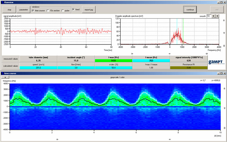
Screenshot of FlowView software for Doppler examinations
The software shows the signal processing from the Doppler shift raw signal (screenshot top left) through the Fourier analysis (screenshot top right) to the color-coded Doppler frequency spectrum (screenshot below) and enables the qualitative (pulse shape) and quantitative (average and maximum frequency, signal intensity) evaluation of the measurements.
SCOPE OF DELIVERY:
| Item No. | Designation |
|---|---|
| 50400 | Ultrasound Doppler FlowDop200 |
| 50130 | Centrifugal pump MultiFlow |
| 50435 | Doppler probe |
| 50160 | Arm model |
| 70200 | Ultrasound gel |
ADDITIONAL EXPERIMENTS:
| PHY13 | Ultrasound Doppler effect |
| PHY15 | Flow laws |
| MED05 | Vascular diagnostics with ultrasound (angiology) |
| MED06 | Blood pressure measurement with ultrasound (Doppler occlusion pressure measurement) |