


MED02 Ultrasound Examinations on the Breast Model
Couldn't load pickup availability
Ultrasound Examinations on the Breast Model
Investigation of a realistic breast model with tumors and their localization and size estimation using the B-mode technique
The examination of a realistic breast model with tumors and their localization and size estimation using the B-mode technique provides a typical application of ultrasound in medical diagnostics.
Keywords: reflection, scattering, ultrasound imaging, pulse-echo technique, A-mode, B-mode, breast sonography, tumor size
Mamma sonography, i.e. ultrasound examination of the breast, is, alongside mammography (X-ray examination), the most important imaging method for diagnosing benign and malignant changes in breast tissue. It is used in the early detection of breast cancer. The strength of sonography lies in its ability to distinguish between changes consisting of solid tissue and cavities filled with fluid (cysts). This method can be used, for example, to take a tissue sample from the breast in a controlled manner. Immediately before an operation, the ultrasound examination can show the exact location of the finding and thus enable the doctor to carry out a targeted intervention. In the experiment, a realistic breast model is first examined for any pathological changes by palpating it with the fingers. This allows the two tumors contained in it to be found and their approximate location determined. The areas found are then examined with the ultrasound probe in A-scan mode, and suitable device parameters and a suitable alignment of the ultrasound probe are set. Using the settings found, a B-scan of the breast model is recorded along a selected line and analyzed.
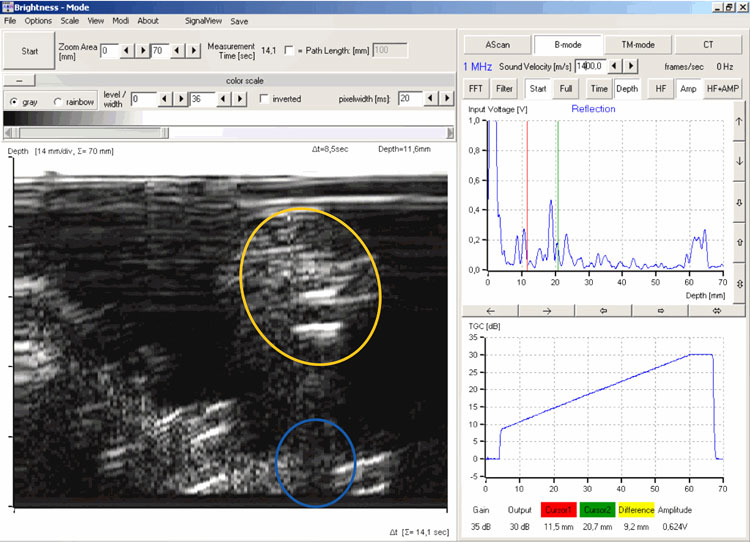
B-mode scan of the first tumor
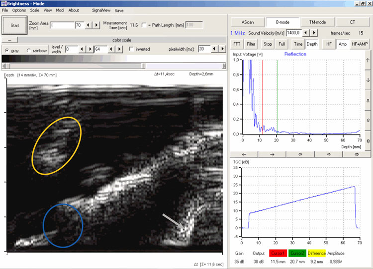
B-mode scan of the second tumor
The ultrasound B-scan images recorded with the measurement software show the tumors with an oval shape and a slightly inclined axis (yellow marking). The attenuation in the tumor tissue is increased, which creates an acoustic shadow on the back wall of the breast model (blue marking).
SCOPE OF DELIVERY:
| Item No. | Designation |
|---|---|
| 10400 | Ultrasonic echoscope GS200 |
| 10151 | Ultrasonic probe 1 MHz |
| 10221 | Breast model |
| 70200 | Ultrasound gel |
ADDITIONAL EXPERIMENTS:
| PHY01 | Basics of ultrasound echography (A-scan) |
| PHY08 | Ultrasound B-scan |
| MED04 | Biometrics on the eye model |
| MED09 | Breast sonography |