


MED01 Ultrasound TM mode (Echocardiography)
Couldn't load pickup availability
Ultrasound TM mode (Echocardiography)
In the experiment, the movement of the heart wall is simulated using a simple heart model. This movement is examined using the time motion method (M-mode). The heart rate and cardiac output are determined using the M-mode recording.
Keywords: Ultrasound echography, reflection, pulse-echo technique, time-motion mode, representation of movement sequences, heart wall movement, echocardiography
In echocardiography, the time motion method, also known as TM mode or M mode for short, is used to examine the movement of the heart and its structures. As with a B-image, the amplitudes of the ultrasound signal echoes of an A-scan are shown on the vertical axis in gray or false color values. The echoes, which are offset in time at a high pulse repetition frequency, are shown next to each other on a horizontal time axis. This creates a curve image that shows the temporal movement of the structure being examined. In the experiment, a movement is generated manually using a membrane. This simulates the periodically repeating movement of a heart wall or heart valve. The measurement software records a TM mode image of the simulated heart wall movement. This can be analyzed and evaluated with regard to the characteristic quantities describing cardiac activity.
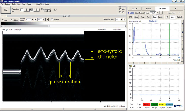
Screenshot of the AScan program in TM mode
The screenshot of the measurement software working in TM mode shows on the left the TM mode recording of a heart wall movement simulated with the simple heart model. The pulse duration and the end-systolic ventricular diameter can be determined from this recording. The heart rate as well as the end-systolic heart volume and the cardiac output can be derived from these two values. In the case of the model, the end-diastolic volume is assumed to be zero.
SCOPE OF DELIVERY:
| Item No. | Designation |
|---|---|
| 10400 | Ultrasonic echoscope GS200 |
| 10154 | Ultrasonic probe 4 MHz |
| 10220 | Heart model |
| 10310 | – optional: tripod set |
ADDITIONAL EXERIMENTS:
| PHY01 | Basics of ultrasound echography (A-scan) |
| PHY08 | Ultrasound B-scan |
| MED03 | Basics of Doppler sonography |
| MED05 | Vascular diagnostics with ultrasound (angiology) |