M33/1 Hand Skeleton Model with Ligaments & Muscles - 3B Smart Anatomy
Couldn't load pickup availability
Hand Skeleton Model with Ligaments & Muscles - 3B Smart Anatomy
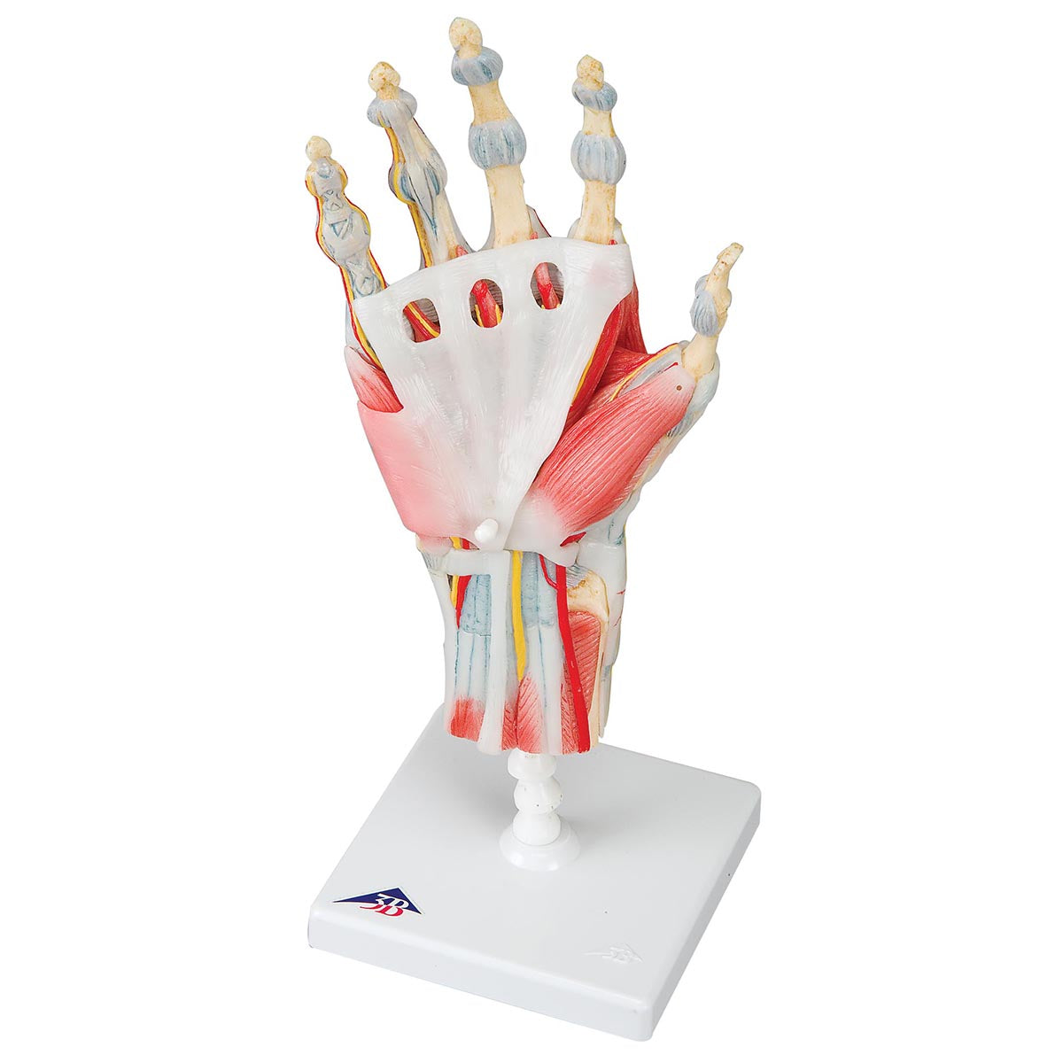
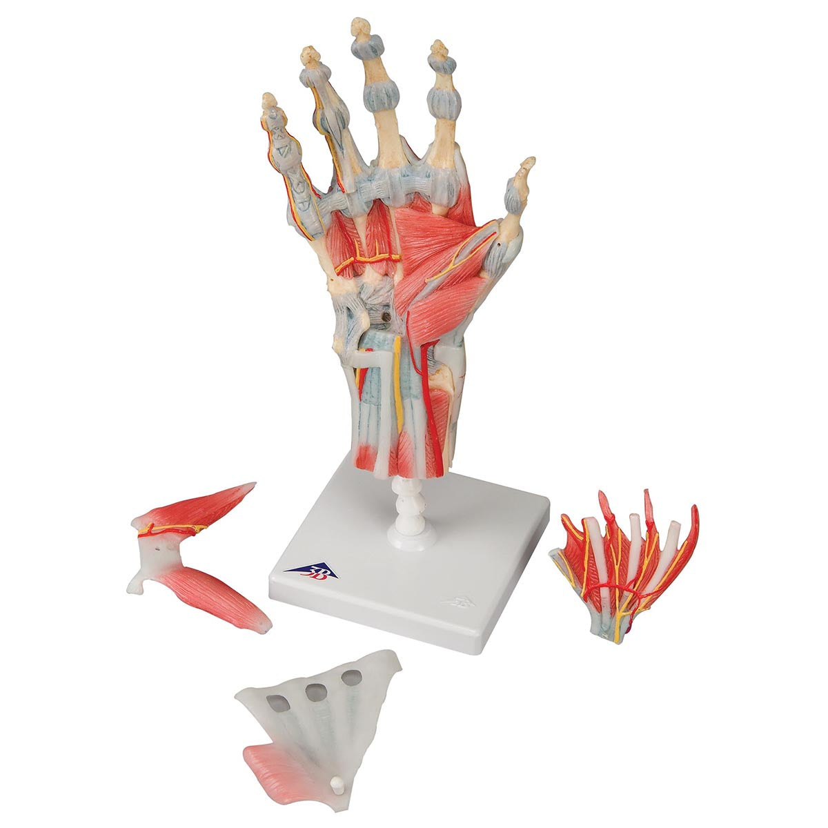
The bones, muscles, tendons, ligaments, nerves, arteries, and veins are all featured in this high quality 4 part model of the hand and lower forearm. The dorsal side of the hand shows the extensor muscles as well as portions of the tendons at the wrist as they pass under the extensor retunaculum. The palmar face of the hand is represented in three layers, the first two are removable to allow detailed study of the deeper anatomical layer of the hand. In addition clinically important structures such as the median nerve and superficial palmar arterial arch can be explored in detail in the hand model. The deepest anatomical layer allows for study of the intrinsic muscles and deep palmar arterial arch in addition to other details of the anatomy of the hand. This high quality anatomically correct hand model with ligaments and muscles is great for detailed study.


